What to Expect at the Diabetic Eye Exam
The eye exam involves various different steps detailed below. It begins with your eye doctor having a conversation with you about your diabetes and overall health. Your vision will be checked. The diabetic eye exam includes pupil dilation and proper checking of the health of your eyes using different kinds of methods and instruments.
Medical History
Your eye doctor will want to hear all about your diabetes and medical background. It is important to share which type of diabetes you have and when you were diagnosed. The eye doctor will ask for details on the medications you take and what you do to manage your diabetes. Any other medical information including eye diseases in first degree relatives is also important to share. Please tell your eyecare provider if you have any eye symptoms or have noticed any changes in your vision.
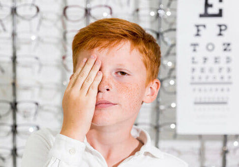
Visual acuity
The eye chart will be used by either your eye doctor or an assistant to check your visual acuity. Your vision can be affected by diabetes and therefore it's important to check how well you're able to see at each visit in order to monitor for any changes. If your clarity of vision decreased, a new prescription for glasses may be given after doing a part of the eye exam known as refraction which determines the best optical correction for you. It’s important to note that the most accurate prescription can be given when your blood sugar has been under control for at least a week prior to your appointment.
Pupil dilation
Pupil dilation is a crucial portion of the diabetic eye exam because it makes the pupils much wider than usual using eye drops. This enables your eye doctor to properly check the back of your eye for any possible damage to the retina, which is the light-sensitive layer of the back of the eye. Diabetic retinopathy is what causes damage to the retina so it's essential for the back of the eye to be properly monitored at each eye exam.
The eye doctor or an assistant will put drops in your eyes to make the pupils larger. This will take effect around twenty minutes after being administered. Usually your pupils will continue to be dilated for approximately two to three hours. We recommend bringing dark sunglasses and to arrange proper transportation for after the appointment to avoid driving with blurry vision due to your dilated pupils.
Fundoscopy
After your pupils have been dilated, the eye doctor will check the back of your eyes and this is known as fundoscopy. There are various signs of diabetic retinopathy and its complications that the eye doctor will look for, such as:
- Abnormality in the blood vessels
- Any type of swelling or blood in the retina
- Fatty deposits
- Formation of some kind of scar tissue
- Growth of new unstable blood vessels
- Detachment of the retina
- Problems with the optic nerve
There are various tools the eye doctor can choose from, such as hand-held instruments or a special microscope, in order to achieve a clear view of the back of your eyes. There is also incredible technology nowadays which allows for a high-resolution photograph to be taken of the back of your eye. This picture can be shown to you on a big screen which allows your eye doctor to show you any changes taking place in your retina. Also, these photographs are stored in your file so that it can be compared with the results at your future appointments.
Fluorescein Angiography
A special dye, called fluorescein, is injected in the arm and travels through the bloodstream to your eyes. A picture is taken in which the fluorescein highlights any abnormalities in your eye’s blood vessels, ensuring an accurate diagnosis.
Optical Coherence Tomography (OCT)
OCT is used for imaging of the back of the eye. It shows the different layers of the retina and any atypical fluid or other abnormalities can be detected. OCT is also used to track the effects of treatment.
Glaucoma testing
Diabetic retinopathy can cause added pressure in your eye which could lead to glaucoma. Your eye doctor may examine your optic nerve during fundoscopy because it is directly related to glaucoma. Also, your eye pressure will be measured in order to screen for this condition.